

Asymmetry is discussed in educational materials about the risk of melanoma as a descriptive feature of certain skin lesions. Changes can include visible differences in structure or outline. Commonly referenced examples involve a dark spot or an irregular mole. Round, evenly shaped moles are typically described as symmetric, whereas lesions in which one side does not mirror the other are described as asymmetric. Asymmetry is presented as an early feature of concern in symptom summaries rather than a definitive diagnosis.

Existing moles may change in shape or size over time. Birthmarks can also be involved. Frequently described locations include the back and extremities, particularly the lower limbs. Some summaries note that patterns can differ between populations, and that fingers and toenail beds may also be affected. Reports mention that lesions can occur on the face and neck, and, less commonly, on mucosal surfaces such as the mouth or rectum.

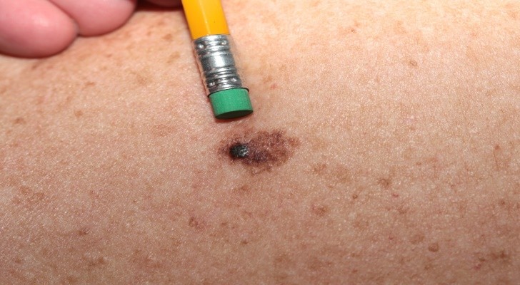
Attention to borders appears in many overviews. Lesions associated with melanoma are often described as having irregular, notched, or blurred edges, in contrast to smooth and regular borders commonly noted in benign moles. Additional surface changes reported in educational sources include bleeding, scaliness, and the appearance of spots or patches around the lesion. Redness or a halo of color and a “foggy” or indistinct outline are also described.

Some individuals with melanoma report episodes of bleeding or local irritation. While many lesions are not painful, symptom descriptions note that sensitivity or tenderness can occur. Accounts also mention itching or sensations described as fluttering or tingling. These features vary widely and are not specific to melanoma but are included in symptom profiles.

A color of the skin change is another frequently cited feature. Educational materials describe benign moles as tending toward a single, uniform shade. In contrast, malignancy-associated lesions may display multiple colors. Common tones include black, brown, and tan, with possible areas of blue, red, or other hues. Changes in color or size are presented as features associated with increased concern in descriptive guides.
The rise of skin relief—such as elevation or thickening—is also described. A lesion that was initially flat may become raised or develop uneven surface contours. Asymmetrical growth patterns are mentioned in summaries, and comparisons with the surrounding skin surface are often used to illustrate the difference.

Another commonly referenced factor is the diameter of the skin’s moles. Educational materials often note that many benign moles are small and uniform in appearance, whereas lesions larger than about six millimeters across raise more concern in symptom checklists. Additional features described include progressive surface disruption, peeling, or scaling. Changes over time—sometimes called “evolution”—are emphasized in summaries that catalog melanoma-associated signs.

Melanoma may be present in the eye (ocular melanoma), which has distinct characteristics. Reports commonly describe involvement of one eye, with features such as light sensitivity or visual changes. Educational resources also note that melanoma can spread to other parts of the body. Discussions of background risk sometimes mention lighter eye color and light skin as factors associated with increased risk in population studies.

Findings in the mouth are described separately as oral melanoma. Summaries note that lumps or swelling can occur on the tongue and may involve lips, teeth, or gums. Red or white patches on the oral mucosa are included among reported features in educational materials. Oral hygiene practices are not presented as predictors in these descriptions; instead, the focus is on recognizing unusual mucosal changes.
Learn more about melanoma and skin cancer at National Cancer Institute.